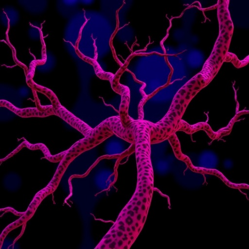
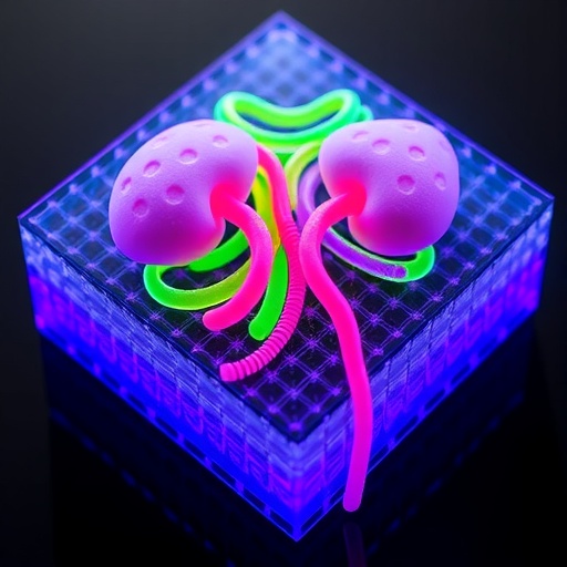
Kidney organoids grown from human stem cells have transformed how researchers study kidney development and disease, but they have always shared one stubborn flaw: they lack blood vessels. Now a team at Maastricht University in the Netherlands has unveiled a 3D printed microfluidic chip that coaxes lab-grown kidney organoids to develop their own primitive capillary networks, both on their surface and deep within their interior, a breakthrough that could push these miniature organs closer to clinical relevance.
The research, published in the journal Biomedical Microdevices, tackles what many consider the single greatest limitation of organoid technology. Kidney organoids derived from human induced pluripotent stem cells, or hiPSCs, can recapitulate the early stages of human kidney development with remarkable fidelity, forming structures that resemble nephrons, the functional filtering units of the kidney. Yet without vasculature, these cellular assemblies remain stunted. The kidneys are among the most richly perfused organs in the human body, receiving between 20 and 25 percent of cardiac output, and tissue more than 100 to 200 micrometers thick simply cannot survive on oxygen and nutrient diffusion alone. Inside conventional organoid cultures, the glomerular rudiments that should eventually form blood-filtering capillary tufts typically remain avascular and disorganized, and prior studies have suggested that the absence of blood flow may even cause the regression of any endothelium that does initially appear.
The Maastricht team, led by Gabriele Addario, Chiara Formica, Lorenzo Moroni and Carlos Mota of the MERLN Institute for Technology-Inspired Regenerative Medicine, approached the problem with a hybrid strategy combining biofabrication, organoids and tissue-derived hydrogels. Their starting point was a deceptively simple manufacturing trick: sacrificial 3D printing. Rather than trying to carve channels out of a solid block, the researchers extruded thin fibers of pluronic F-127, a temperature-responsive polymer that is solid at room temperature but fluid when cold, using a nitrogen-pressure-driven bioprinter fitted with a 250-micrometer nozzle. The fibers were printed at 310 kilopascals onto a base of polydimethylsiloxane, or PDMS, a flexible silicone widely used in microfluidics. A second layer of PDMS was then pipetted over the printed template and cured overnight at 40 degrees Celsius. When the assembly was rinsed with cold phosphate-buffered saline, the pluronic dissolved away, leaving behind two hollow channels with perfectly circular cross-sections and three separate central compartments for gel, all within a fully enclosed chip.
The circular channel geometry is more than an aesthetic choice. Native capillaries are round, and the sacrificial printing approach produces this shape in a way that alternative microfabrication methods, such as stereolithography, cannot easily match. The entire manufacturing process is automated and inexpensive, which the researchers argue is essential if such platforms are to be adopted widely for drug screening.
Once the chip was fabricated, the team seeded human umbilical vein endothelial cells, or HUVECs, into the two channels, where the cells proliferated over seven days until they formed a confluent lining covering roughly 79 percent of the chip’s interior surface. Before that lining could be put to work, however, the researchers had to solve a mundane but critical problem: what to feed it. HUVECs normally thrive in endothelial growth medium, while kidney organoids require a completely different cocktail, typically Advanced RPMI supplemented with glutamine after the organoids reach a certain differentiation stage. A metabolic activity screen using a resazurin-based assay revealed that HUVECs maintained their endothelial identity, as confirmed by staining for the vessel marker CD31, even when grown in the organoid’s own Advanced RPMI medium. The 50:50 mixture of the two media, by contrast, produced the lowest metabolic activity and was abandoned.
The organoids themselves were generated through a carefully choreographed differentiation protocol. The team coaxed hiPSCs separately into two embryonic kidney progenitor populations: metanephric mesenchyme cells, which give rise to nephrons, and ureteric bud cells, which form the collecting duct system. From day 4 of differentiation onward, the two progenitor populations were combined at a one-to-one ratio in low-attachment U-bottom plates, where they self-organized into three-dimensional organoids under the influence of precisely timed growth factors including activin A, FGF9, GDNF and the BMP inhibitor LDN-193,189. By day 14, the resulting organoids contained recognizable nephron-like elements.
The pivotal innovation came with how those organoids were then embedded. In one of the chip’s three central gel compartments, organoids were suspended in a hydrogel made from partially digested decellularized extracellular matrix, or ddECM, derived from pig kidney cortex. A second compartment held organoids in the commercial matrix geltrex, and a third held organoids without any gel at all as a control. The gel-free organoids were literally washed away within days, and the geltrex-embedded organoids progressively lost their three-dimensional architecture. Only the ddECM hydrogel kept the organoids structurally intact while preserving, and even enhancing, their kidney identity. Immunostaining revealed proximal tubule structures marked by the enzyme LTL and distal tubule structures marked by the transcription factor GATA3, with confocal microscopy showing continuous connections between these tubular segments running through the organoid volume.
Proteomic analysis of the ddECM helped explain its potency. Mass spectrometry identified a suite of proteins associated with endothelial cell adhesion, migration and angiogenesis, including fibronectin and its receptor integrin beta-1, laminin subunits, vimentin and moesin. Vimentin in particular has been shown in earlier studies to mimic the activity of vascular endothelial growth factor, the master angiogenic signal, promoting both cell migration and capillary-like tube formation. Moesin, meanwhile, regulates the RhoA/ROCK signaling pathway, a critical mediator of new blood vessel sprouting. Consistent with this molecular profile, organoids embedded in ddECM showed significant upregulation of the angiogenic gene VEGFA alongside markers of epithelial, mesenchymal and podocyte cell populations, indicating that the hydrogel supported vascular development without sacrificing the organoids’ renal character.
When day-14 organoids embedded in ddECM were placed into the pre-endothelialized chip and co-cultured for five days under gentle perfusion, delivered by a rocking platform oscillating five times per minute, the results were striking. The endothelial monolayer lining the channels began to remodel, sprouting branching structures that extended toward the embedded organoids. Primitive capillary-like structures, marked by CD31, appeared not only at the organoid periphery but within its interior, invading regions that had previously been unreachable in static culture. Crucially, these nascent vessels colocalized with LTL-positive proximal tubule-like areas and PODXL-positive glomerulus-like regions, the latter being the very structures that in a real kidney house the blood-filtering capillary tuft. In control experiments, organoids cultured in ordinary U-well plates showed no branching whatsoever, and endothelialized chips maintained without organoids actually lost endothelial coverage, dropping to about 39 percent, underscoring that the organoid-hydrogel combination is what actively recruits and stabilizes the new vasculature.
The achievement represents a meaningful advance over earlier organ-on-chip attempts. A commercially available chip used in prior work placed organoids in a chamber above rectangular channels, resulting in vascularization that was largely restricted to the organoid’s outer edge. The Maastricht chip’s design, with organoids embedded directly in gel compartments flanked by circular vessels, appears to drive vascular ingenuity far deeper into the tissue. The three-compartment layout also allows multiple hydrogel formulations to be tested simultaneously within a single chip, a feature the authors note was absent from previous platforms, along with the ability to generate consistent replicates once conditions are optimized.
The broader stakes are considerable. Kidney diseases affect more than 750 million people worldwide, and the field still lacks humanized three-dimensional in vitro models robust enough for reliable drug testing. Mature, vascularized organoids could enable researchers to model chronic kidney disease and kidney fibrosis in human tissue, screen nephrotoxic drugs before they ever reach patients, and perhaps one day contribute to regenerative therapies. By supplying the missing ingredient, a functioning vasculature that grows into, rather than merely around, the organoid, the Maastricht platform moves the field a significant step closer to kidney organoids that behave less like cellular miniatures and more like the organs they are meant to emulate.
Subject of Research: Vascularization of hiPSC-derived kidney organoids using a 3D printed microfluidic chip
Subject of Research: Technology and Engineering
Article Title: 3D printed chip as platform to vascularize hiPSCs-derived kidney organoids
Article References: Addario, G., Formica, C., Moroni, L., & Mota, C. (2026). 3D printed chip as platform to vascularize hiPSCs-derived kidney organoids. Biomedical Microdevices, 28(2), Article 49. https://doi.org/10.1007/s10544-026-00829-7
Image Credits: AI Generated
DOI: 10.1007/s10544-026-00829-7
Keywords: kidney organoids, hiPSCs, 3D printing, microfluidics, vascularization, HUVECs, decellularized extracellular matrix, organ-on-chip, drug testing, disease modeling
Cite Scienmag News
APA
MLA
Chicago
Denise Maddox. (September 6, 2026). 3D-printed chip vascularizes kidney organoids derived from human stem cells. Scienmag. https://scienmag.com/3d-printed-chip-vascularizes-kidney-organoids-derived-from-human-stem-cells/
Denise Maddox. “3D-printed chip vascularizes kidney organoids derived from human stem cells.” Scienmag, 6 September 2026, https://scienmag.com/3d-printed-chip-vascularizes-kidney-organoids-derived-from-human-stem-cells/. Accessed 6 September 2026.
Denise Maddox. “3D-printed chip vascularizes kidney organoids derived from human stem cells.” Scienmag. September 6, 2026. https://scienmag.com/3d-printed-chip-vascularizes-kidney-organoids-derived-from-human-stem-cells/
Copy citation
Download RIS
Tags: 3D-printed microfluidic chip for organoidsadvances in kidney organoid vascularizationadvancing kidney organoid clinical relevancebiofabrication of kidney tissuesbiofabrication of vascular networks in organoidsblood vessel formation in kidney organoidsclinical relevance of vascularized organoidshiPSC-derived kidney modelskidney organoid vascularizationlab-grown kidney developmentlab-grown kidney with blood vesselsmicrofluidic platforms for organ developmentmicrofluidic technology in biomedical researchorganoid perfusion and nutrient deliveryorganoid tissue engineeringorganoid vascularization challenges and solutionsovercoming diffusion limitations in organoidsregenerative medicine for kidney diseasestem cell-derived kidney modelsstem cell-derived kidney tissue engineeringvascular network formation in organoids