Neurosurgeons facing dysmorphic, wide-neck posterior communicating artery aneurysms sometimes need to drill away a small piece of bone at the skull base, the anterior clinoid process, to reach the aneurysm safely. A new anatomical laboratory study has now quantified, in three dimensions, what that drilling actually does to the working space available for microsurgical clipping, and has added an unusual physical visualization to the field: a cast of the surgical corridor made from liquid gallium.
The study, published in Acta Neurochirurgica, was conducted on five formalin-fixed, color latex-injected cadaveric specimens subjected to a pterional approach, the standard route for many aneurysm clippings. Dissection and anterior clinoidectomy were performed under the operating microscope with neuronavigation, and researchers modeled the volume of surgical freedom, or VSF, before and after the bone removal.
To build the 3D models, the team acquired 17 Cartesian coordinates for each dataset to define the boundaries of the surgical corridor. Neuronavigation produced four paired pre- and postclinoidectomy datasets, taken from the left and right sides of two specimens, along with six additional postclinoidectomy datasets, for a total of 14 coordinate sets.
The headline measurement was modest but directionally clear. Mean volume of surgical freedom increased from 48.1 cubic centimeters before clinoidectomy to 54.2 cubic centimeters after it, a gain of roughly six cubic centimeters. With the small number of specimens involved, however, the difference did not reach statistical significance, with a P value of 0.304.
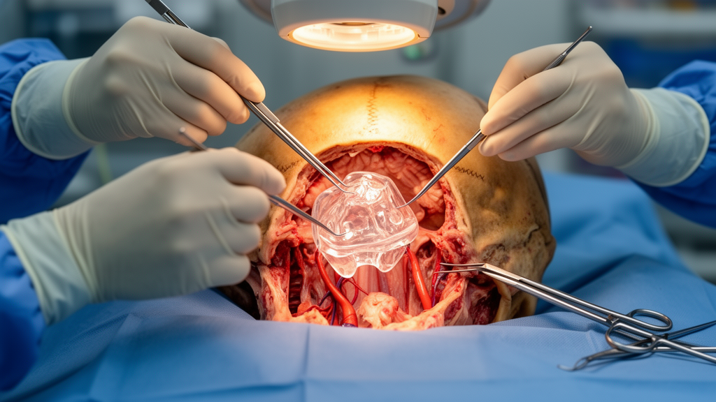
Numbers alone can flatten the anatomy they describe, so the researchers turned to gallium casting as a complementary physical visualization. The molten metal was used to fill the postclinoidectomy surgical space, and the resulting cast gave a tangible representation of the corridor’s true shape, which approximated an irregular two-based pyramid, or frustum. Such casts could serve as a teaching tool, letting trainees hold the geometry of a skull-base corridor in their hands rather than inferring it from a screen.
The clinical context matters. Although many posterior communicating artery aneurysms are increasingly treated endovascularly, the authors note that selected complex aneurysms continue to require microsurgical clipping. For those cases, anterior clinoidectomy is performed to improve exposure, provide proximal control, and increase clip maneuverability, all of which can be decisive when a wide neck resists straightforward clip placement.
The study’s conclusion is deliberately measured. In this cadaveric laboratory model, anterior clinoidectomy was associated with an increase in measured working volume and provided additional operative maneuverability for the clipping corridor. The authors describe these findings as exploratory and frame them as support for the feasibility of 3D VSF modeling and gallium casting as complementary tools for skull-base laboratory training and future preoperative planning research, not as proof of clinical benefit in patients.
That framing is important because the study has clear limitations inherent to its design. Five cadaveric specimens cannot capture the variability of living anatomy, pathological aneurysms, or the physiological conditions of surgery, and the paired datasets came from only two specimens. The lack of statistical significance reflects that small sample size, so the volumetric increase should be read as a consistent trend within this model rather than a definitive effect size.
What the work does offer is a methodological template. By combining coordinate-based volumetric modeling with a physical cast of the surgical space, the approach gives skull-base laboratories a way to compare corridor-altering techniques quantitatively and visually.
Subject of Research: An anatomical laboratory study measuring the effect of anterior clinoidectomy on the surgical corridor for microsurgical clipping of posterior communicating artery aneurysms using 3D volumetric analysis and gallium casting.
Article Title: Novel 3D volumetric and gallium casting assessment of anterior clinoidectomy for microsurgical clipping of posterior communicating artery aneurysms: an anatomical laboratory study
Article References: Morales Roccuzzo, D., Houlihan, L. M., Naughton, D., Bsat, S., Sabahi, M., Gordon, R., Monterroso-Cohen, D., Borghei-Razavi, H., & Adada, B. A. (2026). Novel 3D volumetric and gallium casting assessment of anterior clinoidectomy for microsurgical clipping of posterior communicating artery aneurysms: an anatomical laboratory study. Acta Neurochirurgica. https://doi.org/10.1007/s00701-026-07011-1
Image Credits: AI Generated
DOI: 10.1007/s00701-026-07011-1
Keywords: anterior clinoidectomy, posterior communicating artery aneurysm, microsurgical clipping, volume of surgical freedom, gallium casting, pterional approach, skull base surgery, neuronavigation, cadaveric study, 3D modeling, surgical anatomy, neurosurgery training
Tags: 3D modelinganterior clinoidectomycadaveric studygallium castingmicrosurgical clippingneuronavigationNeurosurgery trainingposterior communicating artery aneurysmpterional approachskull base surgerysurgical anatomyvolume of surgical freedom