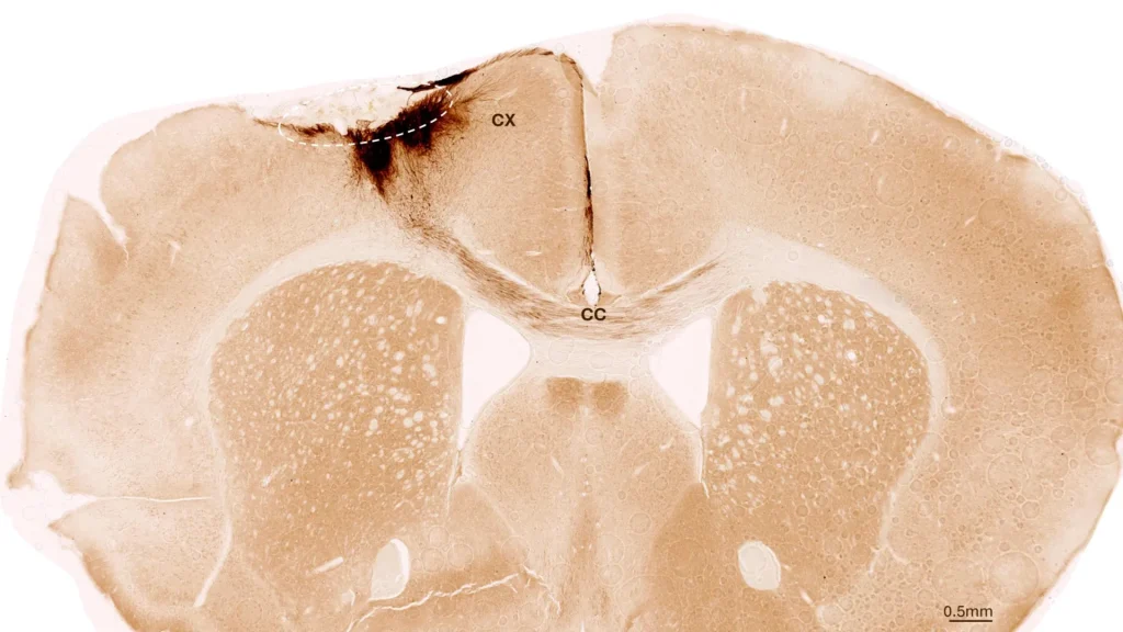
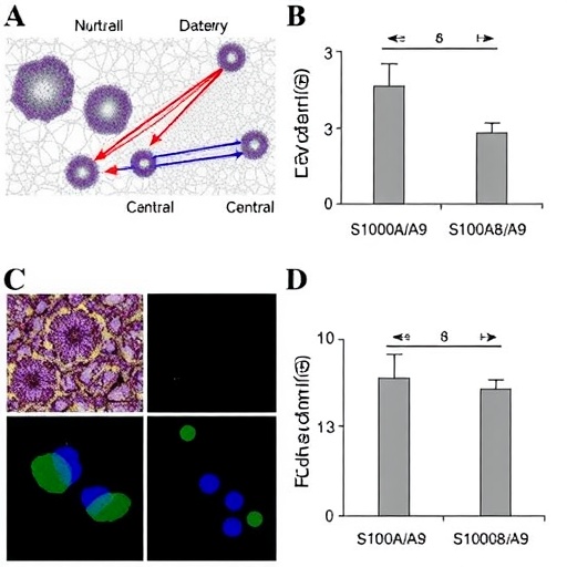
A newly reported immune mechanism may help explain why muscles waste away after losing their nerve supply—and why the body’s own early inflammatory response could also slow that decline. In a study published in Experimental & Molecular Medicine, Xiang, Zhu, Qiu and colleagues describe how neutrophils release the proteins S100A8 and S100A9, which stimulate a reparative population of macrophages and delay atrophy in denervated muscle. The findings place communication between two immune-cell populations at the center of muscle preservation after nerve injury.
Denervation occurs when the connection between a motor neuron and a muscle fiber is disrupted. Without regular electrical and chemical signals from the nervous system, muscle fibers lose contractile activity, alter their metabolism and progressively shrink. This process can follow traumatic nerve damage, spinal cord injury, peripheral neuropathy or certain neurological diseases. Although rehabilitation and surgical repair may restore function in some cases, muscle atrophy can advance before nerve connections are re-established, limiting recovery even when the original injury is treated.
The immune response to denervation is complex. Neutrophils are among the first cells recruited to damaged tissue, where they can destroy microbes, remove cellular debris and release signaling molecules. They are often associated with acute inflammation, but their effects are not exclusively destructive. The new study focuses on a neutrophil population identified by the surface markers CD11b and Ly6G. These cells appear to release S100A8 and S100A9, two calcium-binding proteins that act outside cells as inflammatory and tissue-regulating signals.
S100A8 and S100A9 are frequently produced in response to tissue stress and injury. They can form a protein complex known as calprotectin and influence the behavior of neighboring immune cells through pattern-recognition and inflammatory signaling pathways. In the denervated muscle examined by the researchers, the proteins were linked to the activation of a macrophage state associated with repair rather than prolonged tissue damage. This suggests that signals initially released during inflammation can help redirect the immune environment toward restoration.
The macrophages highlighted in the study were characterized as MerTK-high and Ly6C-low cells. MerTK, or MER receptor tyrosine kinase, is a receptor involved in the recognition and removal of dying cells, a process called efferocytosis. Clearing dead cells is essential because their remains can continue to provoke inflammation if they accumulate. Ly6C-low macrophages are generally associated with a more mature, tissue-supportive phenotype, although macrophage identities exist along a continuum rather than in rigid categories.
According to the researchers, S100A8 and S100A9 released by CD11b-positive, Ly6G-positive neutrophils help activate these MerTK-high, Ly6C-low reparative macrophages. The macrophages may then create a local environment that is more favorable to tissue maintenance, limiting the extent of muscle wasting after denervation. The proposed mechanism does not imply that neutrophils directly restore nerve function. Instead, it indicates that they can influence how muscle responds during the vulnerable period when neural input is absent.
This finding is significant because it challenges a simple view of inflammation as a uniformly harmful force in muscle injury. The timing, intensity and cellular source of inflammatory signals can determine whether they promote damage or recovery. S100A8 and S100A9 are known to participate in inflammatory diseases when their activity becomes excessive or persistent. In the context of denervated muscle, however, the study suggests that a controlled neutrophil-derived signal may support a beneficial transition in macrophage behavior.
The work also offers a possible explanation for why immune-cell composition matters during muscle degeneration. Two tissues with similar levels of nerve damage might undergo different degrees of atrophy if their neutrophils and macrophages respond differently. A treatment designed to preserve or reproduce the S100A8/S100A9 signal could potentially help maintain muscle tissue while nerve repair proceeds. Conversely, indiscriminately blocking these proteins might remove a signal that supports repair, even though suppressing inflammation could appear attractive at first.
Any therapeutic translation will require caution. S100A8 and S100A9 can contribute to harmful inflammation in other settings, and manipulating them systemically could affect infection control, autoimmune activity or cardiovascular health. Future studies will need to define the precise receptors and intracellular pathways through which the proteins influence reparative macrophages, determine how long the signal remains beneficial and establish whether the mechanism operates similarly in different muscles, disease models and human patients. Researchers will also need to distinguish effects on muscle fibers from effects on blood vessels, connective tissue and nerve regeneration.
For now, the study presents denervated muscle as an active immunological environment rather than a passive victim of lost nerve signals. Neutrophils and macrophages appear to participate in a coordinated sequence: early immune cells release S100A8 and S100A9, while a MerTK-high, Ly6C-low macrophage population responds with tissue-supportive activity. By revealing this cellular conversation, the research points toward therapies that do more than suppress inflammation—therapies that could selectively reshape it to preserve muscle until neural function can return.
Subject of Research: Neutrophil–macrophage communication and immune regulation of denervated muscle atrophy
Article Title: S100A8/S100A9 released by CD11b+Ly6G+ neutrophils activate MerTKhiLy6clo reparative macrophages to delay denervated muscle atrophy
Article References: Xiang, Y., Zhu, L., Qiu, Z. et al. “S100A8/S100A9 released by CD11b+Ly6G+ neutrophils activate MerTKhiLy6clo reparative macrophages to delay denervated muscle atrophy.” Experimental & Molecular Medicine (2026). https://doi.org/10.1038/s12276-026-01813-0
Image Credits: AI Generated
DOI: 10.1038/s12276-026-01813-0
Keywords: denervation, muscle atrophy, neutrophils, macrophages, S100A8, S100A9, MerTK, Ly6C, immune regulation, tissue repair