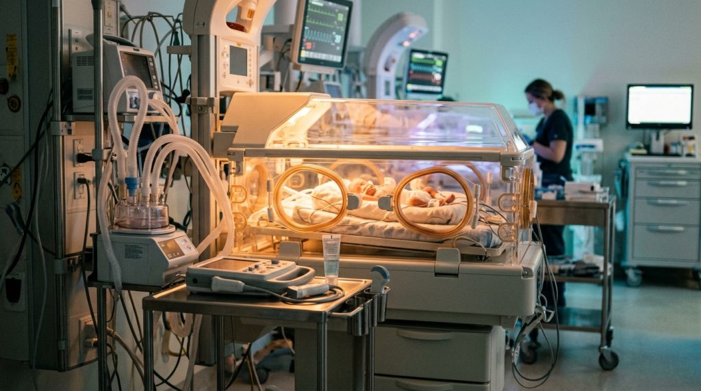
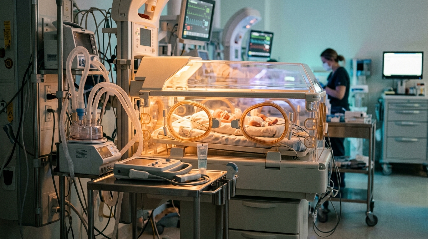
Clinicians have successfully managed a newborn infant suffering from severe Staphylococcus aureus pneumonia complicated by pyopneumothorax and acute respiratory failure by combining ultrasound-guided catheter drainage with high-frequency oscillatory ventilation, according to a case report published in BMC Pediatrics.
Lung ultrasound-guided catheter drainage offers distinct clinical advantages in precise anatomical positioning and dynamic bedside monitoring for neonates, while high-frequency oscillatory ventilation provides established support for severe respiratory failure. Nevertheless, clinical reports describing the concurrent application of both techniques in newborn infants presenting with S. aureus pyopneumothorax and acute respiratory failure have remained relatively rare. Because neonatal physiological characteristics differ substantially from those of older children and adults, pairing oscillatory ventilation to maintain stable alveolar inflation with ultrasound guidance for accurate drain placement is designed to reduce procedural risks and mitigate barotrauma.
The report details the case of a 10-day-old male infant admitted to the hospital after experiencing crying accompanied by throat rales persisting for 17 hours. Upon clinical examination, the newborn demonstrated bilateral lung rattling sounds, occasional grunting, and mild circumoral cyanosis. Initial laboratory testing indicated marked elevation across inflammatory markers, and blood metagenomic next-generation sequencing confirmed S. aureus as the causative infectious agent.
On the infant’s tenth hospital day, his dyspnoea deteriorated suddenly and showed no clinical improvement despite the delivery of high-flow oxygen through a head hood. Clinically diagnosed with acute respiratory failure, the patient underwent bedside chest radiography and lung ultrasound, which revealed a left pyopneumothorax. Medical staff promptly initiated high-frequency oscillatory ventilation for continuous respiratory support alongside ultrasound-guided catheter drainage of the accumulated pleural effusion, and subsequent pleural pus cultures confirmed S. aureus infection.
Following a total hospitalisation course of 32 days, follow-up evaluations demonstrated that the patient’s white blood cell count, C-reactive protein, procalcitonin, and interleukin-6 levels had all returned to normal baseline ranges. The infant’s oxygenation index improved significantly, while chest imaging confirmed that the pulmonary lesion had become more localised, allowing for safe hospital discharge. Continued post-discharge monitoring over a six-month period verified satisfactory physical growth and developmental progress, indicating that this combined respiratory and drainage strategy warrants clinical consideration in severe neonatal presentations.
Subject of Research: Neonatal pyopneumothorax management via ultrasound-guided drainage and high-frequency oscillatory ventilation
Article Title: Ultrasound-guided pleural effusion drainage combined with HFOV in the treatment of a neonatal Staphylococcus aureus pyopneumothorax: a case report
Article References: Zhang, C., Li, W., Bai, Y., Wu, X., Zhang, Y., & Liu, Y. (2026). Ultrasound-guided pleural effusion drainage combined with HFOV in the treatment of a neonatal Staphylococcus aureus pyopneumothorax: a case report. BMC Pediatrics. https://doi.org/10.1186/s12887-026-07482-2
Image Credits: AI Generated
DOI: 10.1186/s12887-026-07482-2
Keywords: neonatology, pyopneumothorax, Staphylococcus aureus, high-frequency oscillatory ventilation, lung ultrasound, pediatric respiratory failure, pleural effusion drainage, Ultrasound-guided, pleural, effusion, drainage, combined
Tags: combineddrainageeffusionHigh-Frequency Oscillatory VentilationLung ultrasoundneonatologypediatric respiratory failurePleuralpleural effusion drainagepyopneumothoraxStaphylococcus aureusUltrasound-guided